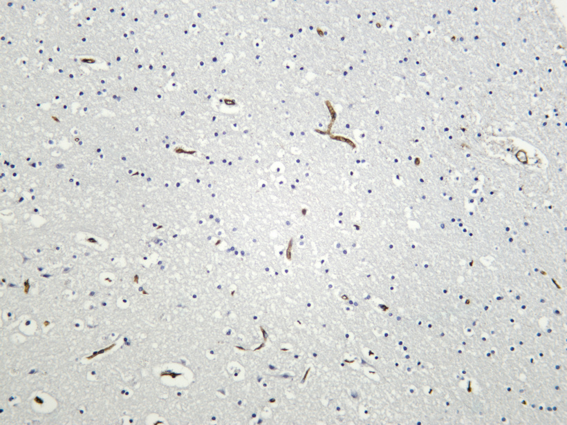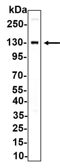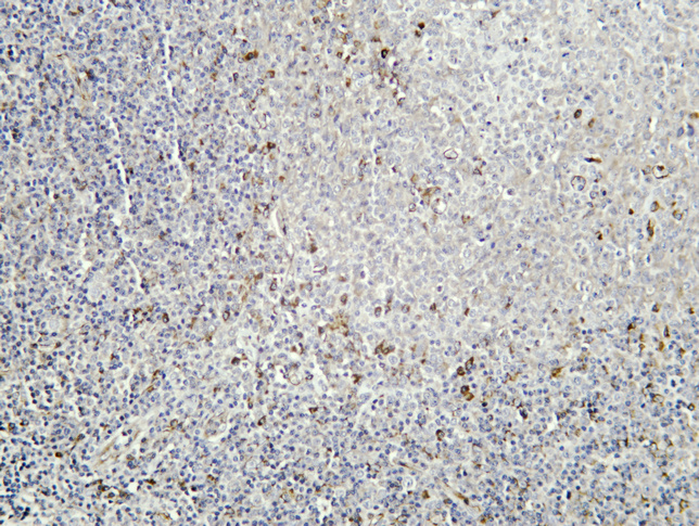

| 别名 | PODXL;PCLP;PCLP1;Podocalyxin;GCTM-2 antigen;Gp200;Podocalyxin-like protein 1;PC;PCLP-1 |
| 基因名称 | PODXL |
| 蛋白名称 | Podocalyxin |
| 推荐应用 | |
| 反应物种 | Human |
| 存储缓冲液 | PBS, 50% glycerol, 0.05% Proclin 300, 0.05%BSA |
| Human Swissprot No. | O00592 |
| Mouse Swissprot No. | Q9R0M4 |
| 特异性 | Endogenous |
| 稀释度 | IHC-P 1:200-1:500;WB 1:2000-1:10000;IF-P/IF-F/IF-ICC 1:200-1:1000;ELISA 1:5000-1:20000;IP 1:50-1:200; |
| 参考分子量 | 130kDa |
| 预测分子量 | 59kDa |
| 运输及保存条件 | Biological ice bag transportation. Store at -20°C for at least 12 months(Do not lower than -25°C). Avoid freeze/thaw cycles |
| 宿主 | Rabbit |
| 同种型 | IgG,Kappa |
| 背景介绍 | podocalyxin like(PODXL) Homo sapiens This gene encodes a member of the sialomucin protein family. The encoded protein was originally identified as an important component of glomerular podocytes. Podocytes are highly differentiated epithelial cells with interdigitating foot processes covering the outer aspect of the glomerular basement membrane. Other biological activities of the encoded protein include: binding in a membrane protein complex with Na+/H+ exchanger regulatory factor to intracellular cytoskeletal elements, playing a role in hematopoetic cell differentiation, and being expressed in vascular endothelium cells and binding to L-selectin. [provided by RefSeq, Jul 2008], |
| 细胞定位 | Apical cell membrane. Cell projection, lamellipodium. Cell projection, filopodium. Cell projection, ruffle. Cell projection, microvillus . Membrane raft . Membrane ; Single-pass type I membrane protein . In single attached epithelial cells is restricted to a preapical pole on the free plasma membrane whereas other apical and basolateral proteins are not yet polarized. Colocalizes with SLC9A3R2 at the apical plasma membrane during epithelial polarization. Colocalizes with SLC9A3R1 at the trans-Golgi network (transiently) and at the apical plasma membrane. Its association with the membrane raft is transient. Colocalizes with actin filaments, EZR and SLC9A3R1 in a punctate pattern at the apical cell surface where microvilli form. Colocalizes with EZR and SLC9A3R2 at the apical cell membrane of glomerular epithelium cells (By similarity). Forms granular, punctuated pattern, forming patches, preferentially adopting a polar distribution, located on the migrating poles of the cell or forming clusters along the terminal ends of filipodia establishing contact with the endothelial cells. Colocalizes with the submembrane actin of lamellipodia, particularly associated with ruffles. Colocalizes with vinculin at protrusions of cells. Colocalizes with ITGB1. Colocalizes with PARD3, PRKCI, EXOC5, OCLN, RAB11A and RAB8A in apical membrane initiation sites (AMIS) during the generation of apical surface and luminogenesis (By similarity). . |
| 功能 | Function:Functions as an antiadhesin that maintains an open filtration pathway between neighboring foot processes in the podocyte by charge repulsion.,PTM:Glycosylated; contains sialic acid.,similarity:Belongs to the podocalyxin family.,tissue specificity:Glomerular epithelium cell (podocyte)., |
| Clonality | Monoclonal |
| 细胞定位 | Apical cell membrane. Cell projection, lamellipodium. Cell projection, filopodium. Cell projection, ruffle. Cell projection, microvillus . Membrane raft . Membrane ; Single-pass type I membrane protein . In single attached epithelial cells is restricted to a preapical pole on the free plasma membrane whereas other apical and basolateral proteins are not yet polarized. Colocalizes with SLC9A3R2 at the apical plasma membrane during epithelial polarization. Colocalizes with SLC9A3R1 at the trans-Golgi network (transiently) and at the apical plasma membrane. Its association with the membrane raft is transient. Colocalizes with actin filaments, EZR and SLC9A3R1 in a punctate pattern at the apical cell surface where microvilli form. Colocalizes with EZR and SLC9A3R2 at the apical cell membrane of glomerular epithelium cells (By similarity). Forms granular, punctuated pattern, forming patches, preferentially adopting a polar distribution, located on the migrating poles of the cell or forming clusters along the terminal ends of filipodia establishing contact with the endothelial cells. Colocalizes with the submembrane actin of lamellipodia, particularly associated with ruffles. Colocalizes with vinculin at protrusions of cells. Colocalizes with ITGB1. Colocalizes with PARD3, PRKCI, EXOC5, OCLN, RAB11A and RAB8A in apical membrane initiation sites (AMIS) during the generation of apical surface and luminogenesis (By similarity). . |


周一至周日 8:00-18:00