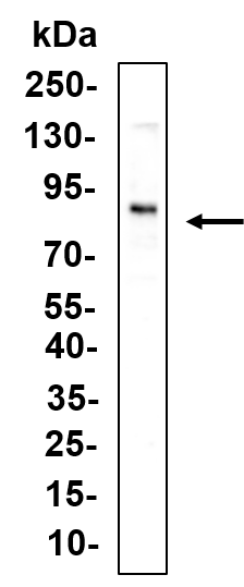

| 别名 | villin 1;VIL |
| 基因名称 | VIL1 |
| 蛋白名称 | Villin-1 |
| 推荐应用 | |
| 反应物种 | Human |
| 存储缓冲液 | PBS, 50% glycerol, 0.05% Proclin 300, 0.05%BSA |
| Human Swissprot No. | P09327 |
| Mouse Gene ID | 22349 |
| Mouse Swissprot No. | Q62468 |
| 特异性 | Endogenous |
| 稀释度 | IHC-P 1:200-1:1000, WB 1:2000-1:10000, IF 1:200-1:1000, ELISA 1:5000-1:20000 |
| 参考分子量 | 93kDa |
| 预测分子量 | 93kDa |
| 运输及保存条件 | Biological ice bag transportation. Store at -20°C for at least 12 months(Do not lower than -25°C). Avoid freeze/thaw cycles |
| 宿主 | Rabbit |
| 同种型 | IgG,Kappa |
| 背景介绍 | This gene encodes a member of a family of calcium-regulated actin-binding proteins. This protein represents a dominant part of the brush border cytoskeleton which functions in the capping, severing, and bundling of actin filaments. Two mRNAs of 2.7 kb and 3.5 kb have been observed; they result from utilization of alternate poly-adenylation signals present in the terminal exon. [provided by RefSeq, Jul 2008], |
| 细胞定位 | Cytoplasm, cytoskeleton. Cell projection, lamellipodium. Cell projection, ruffle. Cell projection, microvillus. Cell projection, filopodium tip . Cell projection, filopodium . Relocalized in the tip of cellular protrusions and filipodial extensions upon infection with S.flexneri in primary intestinal epithelial cells (IEC) and in the tail-like structures forming the actin comets of S.flexneri. Redistributed to the leading edge of hepatocyte growth factor (HGF)-induced lamellipodia (By similarity). Rapidly redistributed to ruffles and lamellipodia structures in response to autotaxin, lysophosphatidic acid (LPA) and epidermal growth factor (EGF) treatment. . |
| Clonality | Monoclonal |
| 细胞定位 | Cytoplasm, cytoskeleton. Cell projection, lamellipodium. Cell projection, ruffle. Cell projection, microvillus. Cell projection, filopodium tip . Cell projection, filopodium . Relocalized in the tip of cellular protrusions and filipodial extensions upon infection with S.flexneri in primary intestinal epithelial cells (IEC) and in the tail-like structures forming the actin comets of S.flexneri. Redistributed to the leading edge of hepatocyte growth factor (HGF)-induced lamellipodia (By similarity). Rapidly redistributed to ruffles and lamellipodia structures in response to autotaxin, lysophosphatidic acid (LPA) and epidermal growth factor (EGF) treatment. . |


周一至周日 8:00-18:00